Craniomaxillofacial surgery follows a multidisciplinary approach, where in many specialists like Craniofacial surgeons, Plastic surgeons, Dermatologist, & Psychiatrist, work together to get best possible cosmetic & functional results so that individuals will get a pleasing & attractive face.
What is Cleft Lip?
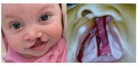
A cleft lip is an opening or split in the upper lip.
What is Cleft palate?
A Cleft palate is an opening or split in the roof of the mouth.
A baby can be born with one or both defects.
Affected children have a range of functional as well as aesthetic problem.
What are the problems these babies face when they are born with such defects ?
Babies with this defect have feeding difficulties at birth due to split in the lip.
They cannot suck the mother’s milk, have swallowing difficulties due to cleft palate and also feeds
comes out through nose, apart from these babies might have Hearing difficulties and Speech
Difficulties too.
Do you know Cleft requires multiple stage of treatment involving many specialist
- 1] 0-2 weeks -Paediatric consultation, Parent counselling, Feeding instructions.
- 2] 2-10 weeks- (infants Orthopaedics) This treatment aims at giving shape to the nose right from the birth and moulding cleft segments which makes surgeon’s job easier and outcome of the surgery will be more precise and natural appearing lip.
- 3] 5-6 months- Surgical repair of the lip.
- 4] 12-15 months-Team evaluation and surgical repair of cleft palate doing surgery at this age will have high impact on development of ideal speech. If delayed child may not develop good speech.
- 5] 3-6 years-team evaluation involving paediatrician,speech therapist, ENT surgeon, Cleft surgeon & Child psychologist.
- 6] 9-11 years - (Alveolar bone grafting) Alveolar Bone graft helps in Eruption of tooth into oral cavity and in turn helps Orthodontist to align teeth in proper shape.
- 7] 12-15 years - Orthodontic treatment.
- 8] 15-18 years - After the orthodontic treatment placement of implants and fixing of bridge, etc, for the missing teeth is done.
- 9] 18-21 years - (Corrective jaw surgery) Surgical advancement of maxilla/bi jaw surgery / genioplasty is done if required.
- 10] 21 years - and above - Lip revision & rhinoplasty.
Infant Orthopedics
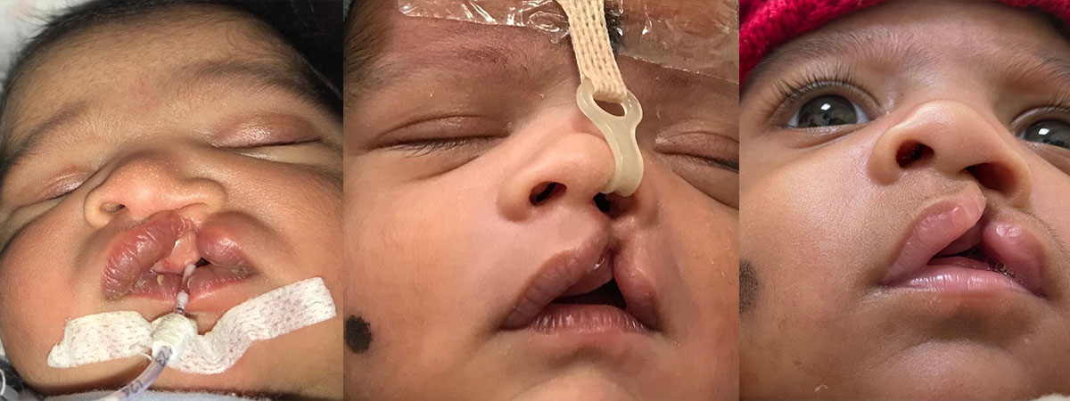
The treatment should be started right from the birth soon after mother discharged from the hospital when baby is 3-4 days old during this period maternal Oestrogen levels are high in babies which will help in moulding the nose and lip to proper shape before actual surgical correction. As the child grows oestrogen levels comes down and moulding capability will be lowered down. It should be done before baby turns 3-4 months age after 4months this treatment is not effective.
Lip Repair
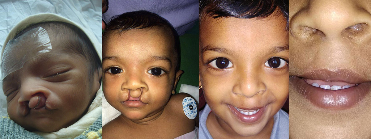
Lip repair is surgically shaping of the lip, and should be done when baby is 5-6 months of age and the hemoglobin should be 10gms. After suture removal, The baby’s lip should be massaged with the prescribes the ointment for a year.
Massaging will help to get proper shape and maintain the shape till it heals, which will give more pleasing lip after a year of surgery.
Palate Repair
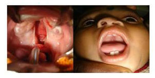
Cleft plate presents serious threat to babies as the opening in the roof of the mouth causes serious chest infection and babies will have difficulty in feeding and can also lead to malnutrition.
Palate closure should be done when baby is 15-18 months of age. This is considered as ideal period during which babies develop speech.
3 months after surgery babies requires team evaluation it’s team of specialist involving speech therapist, paediatric, neurologist, ENT, Psychiatrist to assess normal growth pattern and treatment if required.
Alveolar Bone Grafting
Alveolar bone graft helps in eruption of tooth into oral cavity and in turn helps Orthodontist to align teeth in proper shape.
As the child grows space in the tooth bearing area of gum should be closed.
The graft used is from the hip bone
Dental treatment and Braces therapy
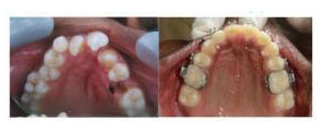
As child grows and teeth begins to erupt it requires attention from paediatric dentist to fill decayed tooth, cap for broken tooth and braces therapy for irregular teeth.
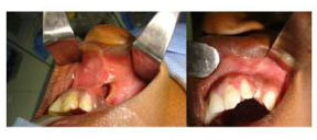
Final Corrective jaw surgery, Cosmetic lip and nose correction
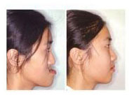
Once Orthodontic treatment is complete corrective jaw surgery is required to get facial balance and pleasing facial appearance.
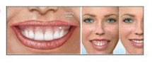
Beauty and Success
Attractiveness is an important part of our personality. Our face is the most important means of communication. A good smile boosts individual confidence levels.
Orthognathic surgery brings you balanced pleasing face with ideal face, jaw & teeth relationship with the help of an operation
What is Orthognathic surgery?
Orthognathic surgery also known as corrective jaw surgery or simply jaw surgery,
Orthognathic surgery is a surgical repositioning of one or both the jaws by means of an operation done from inside the
mouth. Designed to correct abnormal conditions of the jaw and face & its relationship to the teeth
Who requires Orthognathic Surgery?
- Patients with excessive gum show during smiling,
- Small lower jaw,
- Difficulty/inability to eat and talk,
- Forwardly placed upper teeth causing inability to close the lips,
- Big lower jaw, Round face, square face
- Patients with small jaw & associated with snoring (Obstructive sleep apnea)
- Facial and skeletal deformities associated with temporomandibular joint disorder (TMD)
- Birth defect such as Cleft palate , Cleft lip & other associated defect of the face & skull
- Apart from this, people who want to enhance their facial appearance can be considered.
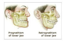
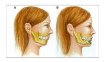
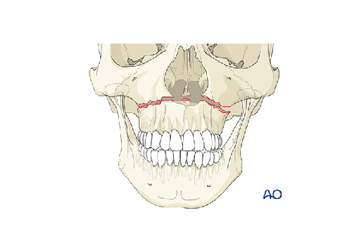
Maxillary & Mandibular Prognathism & Retrognathism
Jaw growth is a slow process that lasts throughout childhood and ends at the young adult age. Sometimes the upper jaw
(maxillary) and lower jaw (mandible) don’t grow at the same rate and the result affects chewing function, appearance,
speech, and oral health.
Mild disproportionate jaw relationship can be corrected by braces alone but some might requires surgical repositioning
of either upper jaw or lower jaw or both along with braces to get harmonious teeth, jaw & face relationship.
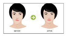
Orthognathic Surgery can correct size and position variations that have affected one or both jaws (maxillary and mandible), among which it can be prognathism (protruding jaw), or retrognathism (retrieving jaw)
Gummy Smile correction
A gummy smile is a smile that reveals excessive gum tissue while talking & laughing which gives unpleasant look to the person’s face a gummy smile occurs when the gums appear to be disproportionately large or prominent when compared with the teeth. The main reason for this is due to over height of the upper jaw (Maxillary bone) which can be corrected by orthognathic surgery and ideal face to teeth relationship can be achieved.
Facial Asymmetry
Facial asymmetry is presence of a clinically significant variation between the two halves of the face that is visible clinically might be due to growth disturbances in upper jaw, lower jaw or both the jaws. Or may be over growth or under growth of mandibular condyle these defects can be corrected by orthognathic surgery.
Malar & Infra orbital Rim Deficiency
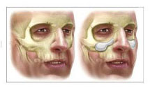
This condition can occur in some patients without any abnormality in the teeth & associated bone structure which is present either from birth or due to loss of bone during previous facial bone injury giving aged appearance and difference in two half’s of the face. This can be corrected either by bone graft or artificial implants specially designed for this purpose which will restore the facial balance & give more pleasing look to the face.
Genioplasty
Also called as Plastic surgery of chin. This surgery is done to make chin look more attractive, either by
advancement, reduction or movement of the chin 3 dimensionally to achieve attractive chin
Patients with small chin, Double chin, large chin and asymmetric chin will benefit by this surgery. Entire
operation is done from inside the mouth so that no visible scars are present from outside.
Double Chin Correction
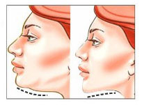
Double chin is a common condition that occurs when a layer of fat forms below your chin this condition is often associated with backwardly placed lower jaw or a small chin or both which can be corrected along with genioplasty & excess of fat can be removed to make the person face more pleasing and acceptable.
how we address face with abnormal size and shape ?
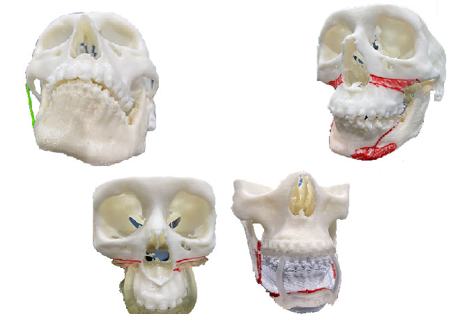
After thorough Clinical examination & CT scan of the facial bones 3D models of the upper & lower jaw is fabricated using latest 3 D printing Technology and then surgery is planned either for upper jaw, lower jaw, chin which will restore the normal jaw & teeth relationship. Post-surgery one may need to wear braces for 6-8moths to get balanced teeth relationship in accordance with newly positioned jaw bones.
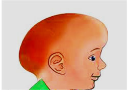
What is Pediatric Craniosynostosis?
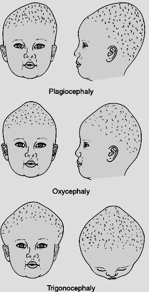
The skull is formed by several separate bones. These skull bones are connected to one another by specialized structures called sutures. These sutures look like seams or spaces between the skull bones. The sutures are growth centers for the skull bones. Craniosynostosis is present when one or more of the sutures closes earlier than it should causing the skull to grow into an abnormal shape..
Babies' brains grow very quickly in the first two years of life. As the brain grows it stretches the sutures which signals the sutures to make new bone. The sutures allow the skull to enlarge and create just enough space for the brain. Normally, these sutures remain open until we reach adulthood, long after the brain and skull have stopped growing. Craniosynostosis causes a baby’s skull to be misshapen because the brain continues to grow at the same rate even if one or more sutures closes too early.
The remaining open sutures have to grow faster to make up for the closed suture. This extra growth causes a change in head shape. In some cases, the remaining open sutures can’t grow fast enough to keep up with the brain’s growth causing an abnormally high pressure in the skull, which can have negative effects on brain health. These include learning delays, blindness, and, rarely, death, if untreated.
What are the different types of Pediatric Craniosynostosis?
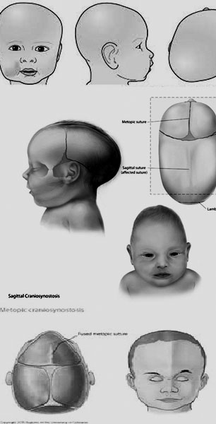
Craniosynostosis can affect babies in two different ways.
1. Isolated craniosynostosis
Isolated craniosynostosis, also known as non-syndromic craniosynostosis, is the closing of only one suture with no other associated health problems and is the most common kind of craniosynostosis.
2. Syndromic craniosynostosis
Syndromes are when three or more medical problems occur in a recognizable pattern. When craniosynostosis is part of a syndrome, it is known as syndromic craniosynostosis. In these cases, there are usually two or more sutures that closed too early. Patients also have other health conditions as part of the syndrome.
What are the signs and symptoms of Pediatric Craniosynostosis?
- Abnormal feeling or disappearing fontanel (soft spot on the top of the head)
- Asymmetrical, misshapen skull
- Development of a raised, hard ridge along the skull
- Slow or no growth of the head as the baby grows
These conditions can be corrected with surgery and can restore normal development of brain & face
The Craniomaxillofacial surgery division provides 24 hour service to attend any emergency following a road traffic accident involving the head & neck region.
- Plastic surgery of cut facial wounds & gums
- Refixing of fallen teeth while playing or due to an accident.
- Fixing of broken jaw bones (Maxilla & mandible)
- Management of secondary traumatic deformity
- Complex facial injury involving eye ,cheek along with brain injury
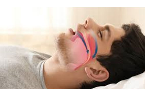
Do you Snore?
What is snoring?
Vibration of respiratory structures and the resulting sound due to obstructed air movement during breathing while sleeping.
What are the causes of Snoring?
- Throat weakness, causing the throat to close during sleep
- Mal positioned jaw, often caused by tension in the muscles.
- Obesity that has caused fat to gather in and around the throat
- Obstruction in the nasal passageway.
- Obstructive sleep apnea
- Sleep deprivation.
- Sleeping on one's back, which may result in the tongue dropping to the back of the mouth
Will it affect our normal Life Style?
Yes In some cases, the sound may be soft, but in most cases, it can be loud and unpleasant. Snoring during sleep may be a sign, or first alarm, of obstructive sleep apnea. Though snoring is often considered a minor affliction, snorers can sometimes suffer severe impairment of lifestyle. Significant improvement in marital relations after snoring was surgically corrected. New studies associate loud "snoring" with the development of carotid artery atherosclerosis. Snoring vibrations are transmitted to the carotid artery, identifying a possible mechanism for snoring-associated carotid artery damage and atherosclerotic plaque development and consequently ischemic stroke.
What are signs of snoring?
- Snoring is known to cause sleep deprivation to snorers and those around them,
- drowsiness,
- Irritability,
- Lack of focus and decreased sexual desire.
- It has also been suggested that it can cause significant psychological and social damage to sufferers.
What happens during Snoring?
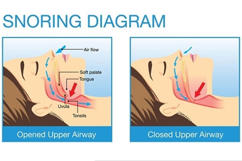
Snoring is caused by a partially closed upper airway (the nose and throat). When this happens, it means that a person isn’t taking in enough oxygen for the body to perform its important functions. The brain then sends a signal to the body to wake up to get the oxygen it needs, likely resulting in the person waking up throughout the night without realizing it Closure of upper airway may be due to blockage of air flow when air enters lungs. Obstruction may be present in the nose, upper jaw, Lower jaw or involving all the structures.
Can it be corrected?
Yes snoring can be treated by clearing the blockage of airflow into the lungs. Clinically expert examines and identifies where the obstruction is and treat accordingly. Craniomaxillofacial surgeon, ENT surgeon, Pulmonologist are involved in treating snoring.
Modes of treatment
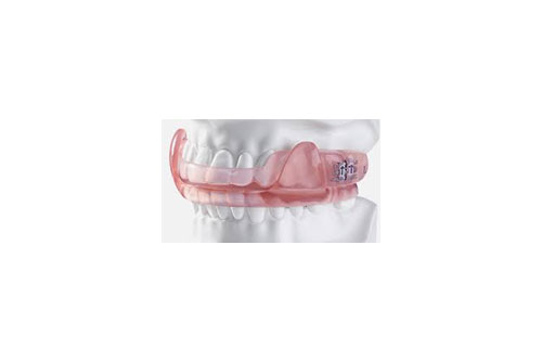
Dental appliances
Specially made dental appliances called mandibular advancement splints, which advance the lower jaw slightly and thereby pull the tongue forward, are a common mode of treatment for snoring. Such appliances have been proven to be effective in reducing snoring and sleep apnea in cases where the apnea is mild to moderate.
Positive airway pressure
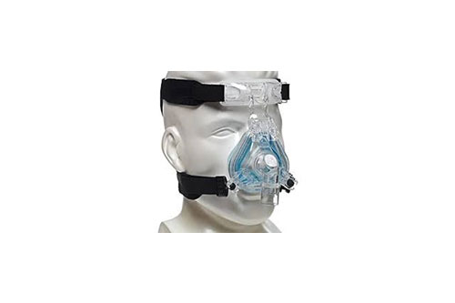
A continuous positive airway pressure (CPAP) machine is often used to control sleep apnea and the snoring associated with it. To keep the airway open, a device pumps a controlled stream of air through a flexible hose to a mask worn over the nose, mouth, or both. This is one of the oldest technique of treating Snoring.
Surgery
Surgical treatments to modify airway anatomy, known as sleep surgery, are varied and must be tailored to the specific airway obstruction needs of a patient.
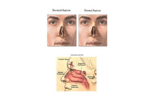
- Nasal surgery, including turbinectomy (removal or reduction of a nasal turbinate), or straightening of the nasal septum, in patients with nasal obstruction or congestion which reduces airway pressure and complicates OSA.
- Tonsillectomy and/or adenoidectomy in an attempt to increase the size of the airway.
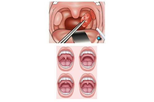
- Removal or reduction of parts of the soft palate and some or all of the uvula, such as uvulopalatopharyngoplasty (UPPP) or laser-assisted uvulopalatoplasty (LAUP).
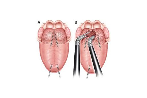
- Reduction of the tongue base for those who has large tongue obstructing airway
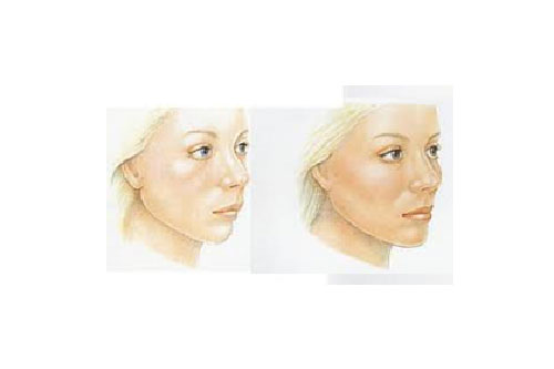
Genioglossus advancement / Genioplasty
In which a small portion of the lower jaw that attaches to the tongue is moved forward, to pull the tongue away from the back of the airway. This surgery improves appearance of the face soon after the surgery.
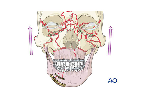
Maxillomandibular advancement
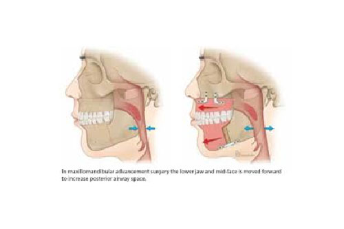
This is the one of the latest treatment for treatment of snoring & obstructive sleep apnoea where either mandible or maxilla or both brought forward to open the airway obstructed by either upper jaw or lower jaw or both.
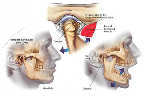
- Treatment for clicking joints.
- Pain on wide mouth opening, joint dislocation, night grinding habits, reduced mouth opening.
- Fractures involving the temporomandibular joint.
- Treatment of locked jaw
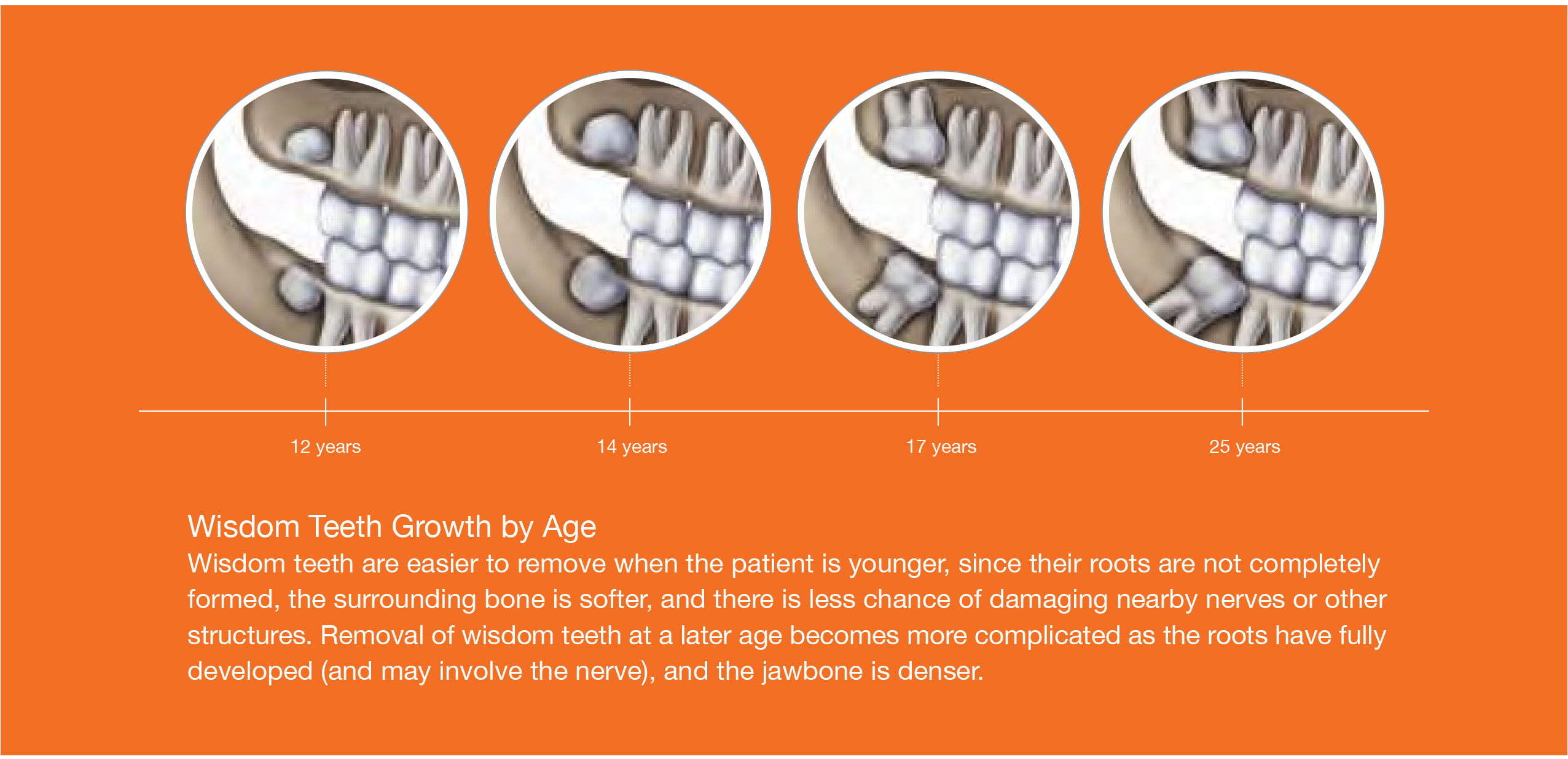
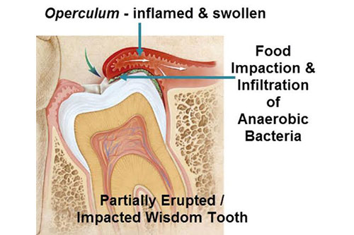
Wisdom teeth, or third molars, are the last teeth to develop and appear in your mouth. They come in between the ages of 17 and 25, a time of life that has been called the “Age of Wisdom.”
What Is an Impacted Tooth?
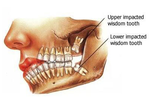
When a tooth is unable to fully enter the mouth, it is said to be “impacted.” In general, impacted teeth are unable to break through the gums because there is not enough room. Nine out of ten people have at least one impacted wisdom tooth.
How Serious Is an Impacted Wisdom Tooth?
If left in the mouth, impacted wisdom teeth may damage neighboring teeth, or become infected. Because the third molar area of the mouth is difficult to clean, it is a site that invites the bacteria that leads to gum disease. Oral bacteria may travel from your mouth through the bloodstream, where it may lead to possible systemic infections and illnesses that affect the heart, kidneys and other organs.
Research has shown that once periodontal disease is established in the third molar areas, the problem is persistent and progressive, but may improve following extraction of the teeth. 4 5 6 In some cases a fluid-filled cyst or tumor may form around the base of the untreated wisdom tooth. As the cyst grows it may lead to more serious problems as it hollows out the jaw and damages surrounding nerves, teeth and other structures.
Must the Tooth Come Out if It Hasn’t Caused Any Problems Yet?
Many people believe that as long as they are not in pain, they do not have to worry about their wisdom teeth. However, pain free does not mean disease or problem free. In fact, wisdom teeth that come in normally may still be prone to disease, according to a study by the American Association of Oral and Maxillofacial Surgeons and the Oral and Maxillofacial Surgery Foundation. AAOMS strongly recommends that third molars be evaluated by an OMS by the time a patient is a young adult in order to assess the presence of third molars, disease status, and to suggest management options ranging from removal to a monitored retention plan to ensure optimal patient-specific outcomes.
- Infections and/or periodontal disease
- Cavities that cannot be restored
- Pathologies such as cysts, and tumors
- Damage to neighboring teeth
In general, dental and medical professionals agree that wisdom teeth should be removed in the following instances:
Wisdom teeth that are completely erupted and functional, painless, cavity-free, in a hygienic environment with healthy gum tissue, and are disease-free may not require extraction. They do, however, require regular, professional cleaning, annual check-ups and periodic radiographs to monitor for any changes.
What Happens During Surgery?
If your dentist or healthcare professional recommends that your wisdom teeth be removed, you will most likely be referred to an OMS for the procedure. Before surgery, your oral surgeon will discuss the procedure with you and tell you what to expect. This is a good time to ask questions. Also talk to your surgeon about any concerns you have. Be sure to let your doctor know about any illness you have and medications you are taking.
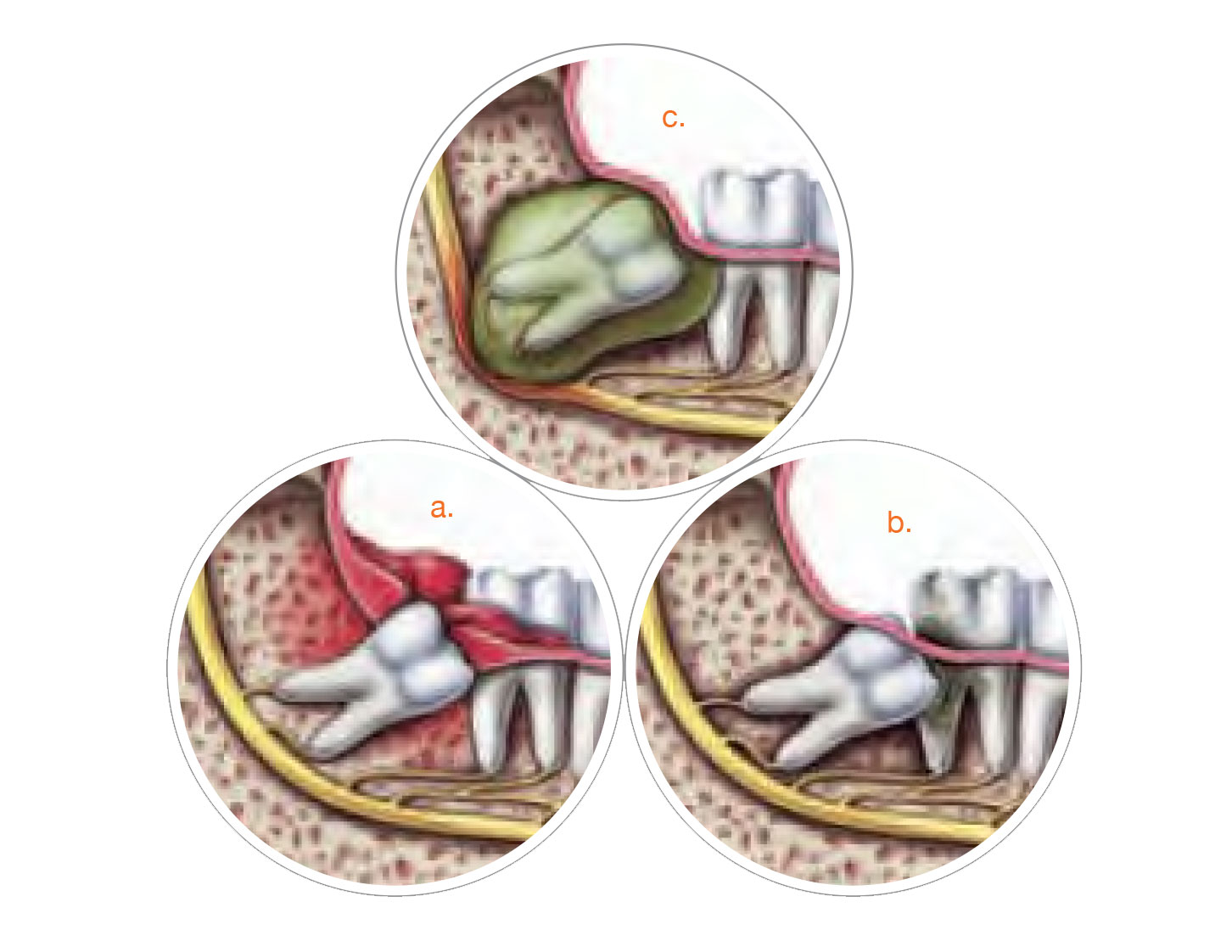
There are several conditions that affect how easy it will be to remove a wisdom tooth. These conditions include how the tooth is positioned and the stage of root development. If the wisdom teeth are impacted the surgery might be more complicated.
Most of the time third molars can be removed with little or no pain. Usually they can be extracted at the oral and maxillofacial surgery office. Patients are given either local anesthesia, intravenous sedation or general anesthesia. Your surgeon will recommend the anesthetic option that is right for you.
What Happens After Surgery?
Following surgery, you may experience some swelling and mild discomfort, which are part of the normal healing process. Cold compresses may help decrease the swelling, and medication prescribed by your Oral and Maxillofacial Surgeon can help manage the discomfort. You may be instructed to modify your diet following surgery and later progress to more normal foods.